🕒 14 min
Co-author: Mario Zelić
“Nothing in life is to be feared, it is only to be understood. Now is the time to understand more, so that we may fear less.”
Marie Curie
Radiation is a word that incites fear in a lot of people. Much of that fear originates from a misunderstanding of what it actually is, what it does and what it does not. “Radiation” is a very broad term, generally used to represent any emission of energy by a source of some kind. However, there are many different phenomena that fall under that umbrella, and they come in varying degrees of rarity and of danger. Even those that may be seen as “dangerous” in some respects can be more useful than harmful – which is why medicine has both treatments for those who suffered too much damaging radiation, and treatments utilizing the purposeful irradiation of a patient.
Again, the word “radiation” can mean a lot of things. In physics, it can usually be split up into two very different (though sometimes related) categories – electromagnetic (EM) radiation and nuclear radiation. Electromagnetic radiation is defined by its effect – it consists of waves of pure energy, oscillations in the electromagnetic field. It is the less interesting of the two for our discussion, so we won’t go into too much detail here, but you may want to check out some of the resources linked at the end of the article to learn more. Nuclear radiation is defined by its source – it originates in the atomic nucleus. The nucleus is made of protons and neutrons, held together by one of the four fundamental forces, the strong force. However, some nuclei are inherently unstable, or radioactive. They decay and emit nuclear radiation to become more stable.
The good, the bad and the ugly
When we talk about radioactivity (and, more generally, any kind of radiation), it is worth noting that it isn’t an either-or type of thing. Things aren’t just radioactive or not, they can be more, less or effectively not at all radioactive. It is the amount and type of radiation that matter. This is why a radiologist can safely expose you to a controlled dose of radiation – the potential harm is so minimal that it is outweighed by the benefits of getting more information towards a diagnosis for you. That is, however, also why they don’t stand in the room with you, or why they wear shielding and you don’t (or why you wear less of it, anyway). A radiologist works with radiation all the time, so they would put themselves at risk if they didn’t minimize their exposure per patient.
An important distinction must be made between different types of radiation. Firstly, we can differentiate between ionizing and non-ionizing radiation. Ionizing radiation has sufficient energy to produce ions in matter at the molecular level, while non-ionizing does not. Examples of non-ionizing radiation can be seen in our everyday life, from our smartphones, radio antennas, visible light and even 5G towers (about which there has been a lot of controversy because of a lack of understanding of what radiation really is). To remain completely honest, it is necessary to point out that non-ionizing radiation can cause some damage to us, but only in the form of heat. This heating effect is also used in medicine today in various forms of clinical infrared and microwave therapies for rehabilitation from surgery and injury, pain relief and much more.
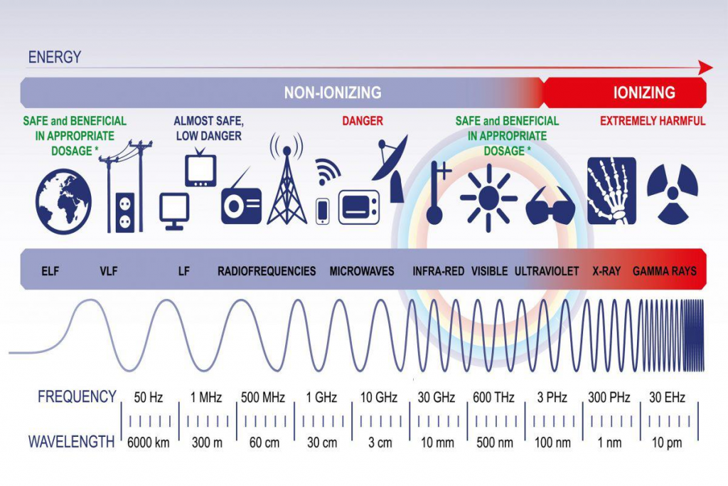
When it comes to ionizing radiation, those ions can do significant damage to DNA and proteins in our cells, which can lead to cell death and various injuries. When we think of ionizing radiation, we usually think of X-ray and gamma ray electromagnetic radiation as well as nuclear radiation. Typical radiation injuries are, but not limited to:
- Anemia (loss of production of red blood cells)
- Digestive tract problems (loss of epithelial cells which cover the inside of our gut and help us with digestion)
- Tissue reaction (when large doses of radiation damage large portions of tissue such as our skin, lungs, blood vessels and brain)
- Mutation of sperm and egg cells (slightly increased risk of genetic defects in children)
- Increased chance of developing cancer
These radiation injuries commonly present themselves in three stages:
- First (or acute = rapid onset) stage – nausea, loss of appetite, vomiting, tiredness and diarrhea
- Second (or latent = waiting) stage – no symptoms
- Third (or chronic = long-term) stage – various syndromes depending on the absorbed dose
The severity of damaging effects of radiation depends on many factors, but the most important ones are:
- The amount (dose)
- The time the dose is received
- How much of the body is exposed
- Sensitivity of the tissue to radiation
- Person’s age, health, presence of genetic abnormalities etc.
When it comes to nuclear radiation, three types are most commonly distinguished, and those are alpha, beta and gamma radiation, depending on the particles emitted.
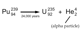
Alpha radiation comes from the term “alpha decay” in nuclear physics, where an unstable atomic nucleus breaks apart into an alpha particle (2 protons and 2 neutrons, which you may recognize as a helium-4 nucleus) and a daughter nucleus which has 2 protons and 2 neutrons fewer.
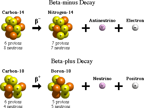
Beta radiation comes from beta decay, in which an unstable nucleus breaks down, releasing a so-called beta particle. There are two types of beta decay, beta-minus and beta-plus decay, with two types of beta particles. In beta-minus decay an unstable nucleus breaks down into an electron, an anti-neutrino (the neutrino’s antiparticle partner) and a daughter nucleus which has one extra proton. In beta-plus decay, an unstable nucleus breaks down into a positron (an electron-like particle which has the same mass, but has a positive charge instead), a neutrino (a small, almost massless particle which has no charge and carries large amounts of energy) and a daughter nucleus which has one proton less. The release of these fast, high-energy electrons or positrons is what we usually refer to as beta radiation.
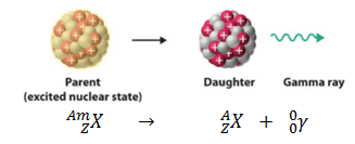
Gamma radiation comes from gamma decay, where an excited nucleus (one with a large amount of energy stored in it) releases energy in the form of a gamma photon (which is really a form of high-energy electromagnetic radiation).
In order to know how much radiation a person has been exposed to, we need a way to measure the amount of the radiation a person has absorbed. This is known as the absorbed dose, and it is a measure of the energy deposited by ionizing radiation. It is equal to the energy deposited per unit mass, so it has a unit of joule (energy) per kilogram (mass), with the adopted name of gray (Gy) where one gray is equal to one joule per kilogram. Sometimes we can also encounter an older unit of absorbed dose called rad where 100 rad = 1 Gy.
A clinically more significant measure of radiation is the equivalent dose. The equivalent dose is equal to the absorbed dose multiplied by a radiation weighting factor. This factor is dependent on the different types and energies of radiation a person is exposed to. For example, this factor is 1 for X-rays, gamma rays and beta particles, 5 for protons, between 5 and 20 for neutrons and alpha particles. The unit for equivalent dose is the Sievert (Sv) and is used to measure how much radiation a person can safely absorb before some of the symptoms start to appear. For example, occupational exposure (meaning how much radiation a radiologist or nuclear medicine specialist can absorb in one year) is 20 mSv. Different parts of the body have different equivalent doses, so our eyes, which are most sensitive to radiation, can have only 150 mSv/year, while our hands and feet can sustain as much as 500 mSv/year before a certain number of cells has been affected by radiation. Fetuses are exceptionally vulnerable to radiation, especially during the first few weeks when most commonly mothers are not even aware that they are pregnant, and their equivalent dose is as low as 1 mSv over the duration of pregnancy.
Radiation and medical imaging
Now, knowing that there are ways to safely use radiation to diagnose diseases, let’s see how people have found different use cases over the years for different types of radiation.
The most famous radiation diagnostic technique is X-ray scanning because of its history, as well as numerous useful applications. We use X-rays to look at the internal organs of the body, as well as the bones and (sometimes) blood vessels.
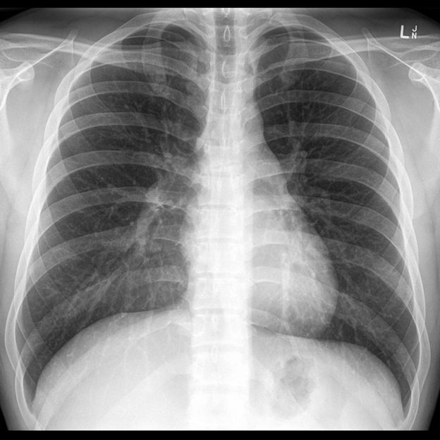
This is a frontal image of a human chest X-ray. Bones are the most visible as they absorb the most radiation, while the lungs are mostly air, which does not absorb that much X-ray radiation, making them appear black on the image. An interesting fact about the X-ray image is that it is a 2D representation of a 3D structure of the human chest. That is why we can see the whole ribs, even though some parts of them are in the front, and some are in the back. In the middle we can see the silhouette of the heart. Around the heart, you can see these light grey net-like structures – those are actually blood and lymph vessels of the lungs, as well as the bronchioles. We can see them in the center since they are big there, before they go through the lungs and become smaller and smaller. This is why we cannot see them at the outer parts of the lungs (unless there is something that makes them visible, like inflammation, a tumor or an accumulation of liquid know as pulmonary edema).
X-ray machines are generally entirely functionally based in electromagnetism, usually made to force fast electrons to collide with heavy atoms, making them release their energy as heat, but also a bit of electromagnetic radiation. X-rays are really high-energy EM radiation, which is why they can pass through your body to form an image on the other side. Regular old light can do something similar in some cases. Placing, for example, your phone’s flashlight directly on the tip of your finger can give you a bit of an inkling into how the bone is positioned inside. It’s basically the same effect – the rest of your body is just too dense for low-energy light to pass through, it gets absorbed or reflected too fast.
As a side note, this ties into why those radiation weighting factors from before are, perhaps confusingly, higher for lower-energy radiation. Your body won’t hold on to a lot of gamma radiation, it will just pass straight through! Alpha particles, on the other hand, are a lot bigger and get stopped more easily. When they stop, they deposit their energy into whatever is around them, causing a lot more damage than if they just went through without stopping. You actually absorb surprisingly little X-ray radiation while getting an X-ray done because of this. Most of your body just isn’t dense enough to stop such high-frequency light.
Next up, you can see what and image of human skeleton scintigraphy looks like.
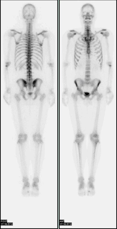
Scintigraphy is a nuclear medicine diagnostic procedure during which a person is injected with a relatively low dose of a radionuclide (an unstable, radioactive atom). Those atoms break down and emit gamma radiation, which is captured by detectors called gamma cameras. The process is similar to X-ray imaging, but it uses higher-energy photons of nuclear origin. Depending on which radionuclide is injected into the patient, different parts of body will absorb the atoms and give off gamma photons.
Scintigraphy is mostly used for looking for various bone diseases such as inflammation, tumors, and fractures. It can, however, be used to diagnose many other organ diseases, for example: gall stones in the biliary system, differentiating benign from malign lumps in the thyroid and parathyroid glands, seeing how a heart functions after a heart attack and a lot more. In the picture above you can see a 2D picture of a human skeleton. The bones which have the highest rate of something going on, like bone metastases or repairing of the bones, will absorb the greatest amount of radiation and show up as the darkest. There are more sophisticated versions of scintigraphy known as SPECT and PET which are becoming more affordable and by that, more commonly used.
Scintigraphy is a specialized use case of a method known as scintillography (both taking their root from the Latin word for “spark”), which refers to any imaging of pulses of radiation released as a result of nuclear decay or certain particle collisions. In physics, it can be used to detect neutrinos, which are notoriously tough to catch.

Another very popular radiology technique is a CT scan. CT stands for computed tomography and is a very sophisticated X-ray machine. By rotating the X-ray source around a person, we can get very detailed look at a particular cross section of a patient’s body. In the GIF, you can see a CT scan of a patient’s abdomen. This patient has stomach cancer which can be seen as a marked abnormal grey structure on the front part of the stomach (front is up, back is down). CT scans are very useful since they can give us a very detailed look of the patient’s organs, blood vessels and it can also show us how organs move and work in real time. This way we can also diagnose functional diseases of various organs.
You may be wondering why we can see much more soft tissue in these scans than we could in the chest X-ray from before. As you know from earlier, softer tissues also stop some of the X-rays from coming through, just not as much as the much harder, denser bones. This is why a CT scanner make use of rotation – and computers (which is where the C in CT comes from). Images taken from different angles can be used to reconstruct a far more complete picture of the various tissues the X-rays passed through, based on their differences in density. The reconstruction process also gets rid of the 3D to 2D ghosting effect, resulting in clean slices. You can see a simple explanation here (or check out a more detailed one at the end of this post).

As the final example of medical imaging, we present to you the queen of radiology – MRI, standing for magnetic resonance imaging. This technique has provided us with immense knowledge about the entire human body, but especially of the human brain. It is commonly used in brain imaging because it can provide a better visual difference between the grey and white matter and the pathological processes happening inside the brain, so it can be used for diagnosing various conditions of the central nervous system such as dementia, multiple sclerosis, Alzheimer’s disease, epilepsy and many other. It is also used in guided stereotactic neurosurgery and radiosurgery for treatment of brain tumors and various malformations. Not to be confused, it is also used for diagnosing diseases of other organ systems, such as the cardiovascular, digestive, and musculoskeletal systems.
Another interesting distinction of MRI as opposed to the other imaging techniques we mentioned is that it does not expose the patient to any ionizing radiation. Instead, MRI scanners produce strong magnetic fields and exploit a very interesting, though also quite complex phenomenon known as nuclear magnetic resonance. The use of strong magnets is also its downfall when it comes to patients with metal implants, however. But, in brief, the nuclei of hydrogen atoms (which are radiology’s atom of choice because they are extremely abundant in the human body, from water to all sorts of organic molecules) can be made to emit signals by absorbing energy while aligning with the strong external magnetic field (because protons exhibit a certain inherent magnetism, their magnetic moment). This is also why, while important, the strength of the outside field isn’t truly the most important factor here – rather, it is the straightness of the magnetic field lines (or the homogeneity of the magnetic field) that does the trick.
Once again, radiation is a word that is both feared and poorly understood by many people. Understanding it, its effects and how it can actually be used to out benefit is crucial in today’s world because, really, various forms of radiation are everywhere in our daily lives. We do, however, hope we have made it a bit more familiar to those of you who needed it, or sparked a deeper interest for those who were already there. So, enjoy its various forms, some of which enable you to read articles such as this one.